BiteSized Immunology: Systems & Processes
T-cell activation
Signal One
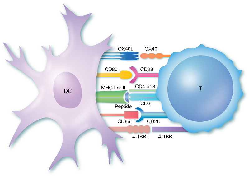
T cells are generated in the Thymus and are programmed to be specific for one particular foreign particle (antigen). Once they leave the thymus, they circulate throughout the body until they recognise their antigen on the surface of antigen presenting cells (APCs). The T cell receptor (TCR) on both CD4+ helper T cells and CD8+ cytotoxic T cells binds to the antigen as it is held in a structure called the MHC complex, on the surface of the APC. This triggers initial activation of the T cells. The CD4 and CD8 molecules then bind to the MHC molecule too, stabilising the whole structure. This initial binding between a T cell specific for one antigen and the antigen-MHC it matches sets the whole response in motion. This normally takes place in the secondary lymphoid organs.
Figure 1. Interaction between T cell and dendritic cell.
Signal Two
In addition to TCR binding to antigen-loaded MHC, both helper T cells and cytotoxic T cells require a number of secondary signals to become activated and respond to the threat. In the case of helper T cells, the first of these is provided by CD28. This molecule on the T cell binds to one of two molecules on the APC – B7.1 (CD80) or B7.2 (CD86) – and initiates T-cell proliferation.
This process leads to the production of many millions of T cells that recognise the antigen. In order to control the response, stimulation of CD28 by B7 induces the production of CTLA-4 (CD152). This molecule competes with CD28 for B7 and so reduces activation signals to the T cell and winds down the immune response. Cytotoxic T cells are less reliant on CD28 for activation but do require signals from other co-stimulatory molecules such as CD70 and 4-1BB (CD137).
T cells must recognise foreign antigen strongly and specifically to mount an effective immune response and those that do are given survival signals by several molecules, including ICOS, 4-1BB and OX40. These molecules are found on the T-cell surface and are stimulated by their respective ligands which are typically found on APCs. Unlike CD28 and the TCR, ICOS, OX40 and 4-1BB are not constitutively expressed on T cells. Likewise, their respective ligands are only expressed on APCs following pathogen recognition. This is important because it ensures T cells are only activated by APCs which have encountered a pathogen and responded. Interaction of the TCR with peptide-MHC in the absence of co-stimulation switches the T cells off, so they do not respond inappropriately.
Signal Three
Once the T cell has received a specific antigen signal and a general signal two, it receives more instructions in the form of cytokines. These determine which type of responder the cell will become – in the case of helper T cells, it will push them into Th1 type (cells exposed to the cytokine IL-12), Th2 (IL-4), or IL-17 (IL-6, IL-23). Each one of these cells performs a specific task in the tissue and in developing further immune responses.
The resulting cell population moves out to the site of the infection or inflammation in order to deal with the pathogen. Other cells present at the tissue site of inflammation– such as neutrophils, mast cells, and epithelial cells – can also release cytokines, chemokines, short peptides and other molecules which induce further activation and proliferation of the T cells.
© The copyright for this work resides with the BSI.
This was updated in 2022