BiteSized Immunology: Experimental Techniques

Multiplex analysis of cytokines
Introduction
The ELISA is a well-established method for quantifying a cytokine of interest in liquid samples. Multiplexing extends this to the measurement of multiple analytes in the same sample. Several multiplexing platforms exist, including bead-based (Luminex and flow cytometry cytokine bead array, CBA) and electrochemiluminescence (carbon surface, MSD) systems.
Assay setup
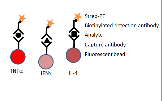
The following description is most relevant to the Luminex, although the other systems are also sandwich immunoassays, using capture and labelled detection antibodies. The principle of this assay is similar to a capture sandwich ELISA, except that the capture antibody is coated onto beads in suspension, rather than being directly coated to the well. Beads are pre-coated with capture monoclonal antibody, specific for a single cytokine, and are internally labelled with a unique combination of fluorescent dyes. By mixing different beads, up to 100 different analytes can be detected simultaneously. Beads specific to the cytokines of interest are incubated with a sample containing the target cytokines in a single well of a 96-well plate. After washing to remove unbound protein, biotinylated detection antibodies (again specific for each cytokine of interest) are added. Streptavidin-Phycoerythrin (Strep-PE), which binds to the biotinylated detection antibodies, is then added. Additional wells are allocated for standard controls containing known amounts of each cytokine.
Samples are acquired on a machine that, like a flow cytometer, uses lasers to simultaneously identify each bead according to its fluorescence signature (corresponding to a single cytokine) and measures the PE signal (the amount of cytokine captured on the bead surface). The PE intensity corresponding to the standard curve of each cytokine is derived simultaneously for each unique bead set. The electrochemiluminescent method consists of a carbon electrode pre-coated with cytokine capture antibodies, with discrete carbon spots relating to each cytokine in the multiplex panel. The sample or standard is then added, followed by an electrochemiluminescent-labelled detection antibody. A voltage is then applied to the plate and the light emitted from the label is quantified.
Example results
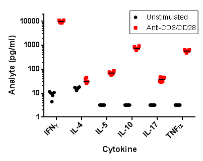
T cells were purified from human blood, and then cultured in the presence or absence of anti-CD3/anti-CD28. After 72 hours in culture, six cytokines were measured in culture supernatants by Luminex. (Although much larger panels are possible, we routinely design bespoke panels of up to eight key markers to ensure that all are within the quantifiable range).
Advantages
Multiplexing is a powerful method generating quantitative data for many analytes from a sample of <50 l; a particular advantage when sample volume is limiting. It can measure multiple markers associated with disease states or mechanism of action studies, expanding our understanding above and beyond the well-established analytes.
Multiplex analysis of cytokines – Bitesized Immunology
© The copyright for this work resides with the author