BiteSized Immunology: Experimental Techniques

Proliferation assay
Objectives of the assay
The in vitro proliferation assay can be used to determine whether or not cells are triggered to divide after exposure to a specific stimulus, or to assess differences between cell populations in their ability to divide in response to the same stimulus.
Procedure
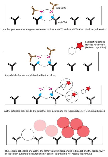
1) Cells in culture are given a specific stimulus. For T cells, this is commonly plate-bound anti-CD3 antibody in combination with soluble anti-CD28 antibody (see figure). Other commonly used compounds to stimulate T-cell proliferation are Con A, PHA, and PMA and ionomycin.
2) A radio-labelled nucleotide is then added to the culture media. The most commonly used is 3H-thymidine. As the cells are stimulated to divide, they utilise the radio-labelled nucleotide in the culture media and incorporate it into their newly-synthesized DNA. Each successive generation of daughter cells will incorporate more of the radio-labelled nucleotide into its DNA.
3) After this incubation (generally 24-48 hours), the cells are removed from the culture media by centrifugation and washed to remove any free radio-labelled nucleotide that has not been incorporated into the cells’ DNA. The total radioactivity is measured, and compared against a control group of cells that did not receive the proliferation-inducing stimulus.
Limitations and alternative approaches
The proliferation assay as described above provides information about the proliferation of a population of cells as a whole, rather than about individual cells. This assay is relatively quick and inexpensive to perform, but does involve the use of radioisotopes, and a specialized machine to read the level of radioactivity present in the samples.
An alternative experimental approach which provides more information about individual cells is to label cells in culture with a fluorescent marker, such as carboxyfluorescein succinamidyl ester (CFSE), which is diluted in each generation of daughter cells as they divide, and can be measured by flow cytometry.
© The copyright for this work resides with the author