BiteSized Immunology: Organs & Tissues

Lymph Node
A number of specialised tissues are important for the proper functioning of the immune system. Among these are the lymph nodes, which provide an ideal environment for communication between immune cells. This environment is necessary for proper activation of the T and B cells (or lymphocytes) that are required for defence against many pathogens. A number of features of lymph nodes help them to perform their functions.
Lymph node location
The lymph nodes are strategically located at locations where they can be easily reached by immune cells travelling around the body. The exact total number of human nodes is not known, but each person is thought to have at least 500. Each lymph node is well-supplied by both lymphatic vessels and blood vessels, which allow lymphocytes to enter and exit. The nodes are contained within a tough capsule, and surrounded by specialised fatty deposits, both of which may give some physical protection.
Lymph node structure
Each human lymph node is up to 20mm in diameter, and is divided into compartments. Each compartment has important functions in enabling communication between lymphocytes. The outer layer (Cortex) contains the B-cell areas, or follicles. The middle layer (Paracortex) is mostly populated by T cells and dendritic cells (Figure 1). The paracortex also contains specialised blood vessels (high endothelial venules) through which many B and T cells enter the node. The lymph vessels enter the nodes at the outer edge, between the capsule and the cortex, and also penetrate deep within the nodes, via channels called conduits. T and B cells leave the node via “efferent” lymphatic vessels, found in the central “medullary” region. All these structures are maintained by a network of fibrous non-lymphoid cells that can also actively influence immune responses.
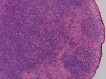
Figure 1. A slice through a lymph node, showing B and T cell areas where lymphocytes are tightly packed (purple).
Co-ordinated movement of cells in lymph nodes
T cells enter the lymph nodes through high endothelial venules, and move around within the T-cell area, transiently interacting with large numbers of dendritic cells. They finally leave the node via the efferent lymphatic vessels. B cells enter by the same route and migrate through the T-cell area to the follicles, before finally leaving the node and re-entering the circulation. These migratory patterns give dendritic cells, T cells, and B cells many opportunities to interact. The appear to travel along the fibrous structural network that supports the node, further increasing their chances of interacting.
Responding to new infections
Lymph nodes are extremely important in responses to infections, especially those that an individual has not previously encountered. Proteins from the infecting micro-organism will reach dendritic cells in the lymph node, or will be carried to the lymph node by migrating dendritic cells. Protein fragments from the organism will be “presented” to T cells. The continual interactions between dendritic cells and T cell ensure that a T cell will soon be found that recognises the infection-derived protein fragment. This T cell will then divide and coordinate the immune response against the infection. Crucially, some of the dividing T cells’ daughters will travel to the B cell follicle and promote B-cell division and maturation, enabling the production of the antibodies that are essential for fighting many infections.
This article was updated by the author in January 2021
© The copyright for this work resides with the BSI.