BiteSized Immunology: Pathogens & Disease

Biofilms and their role in pathogenesis
What is a biofilm?
A common misconception of microbial living is that bacteria exist as individual organisms in a ‘planktonic state’. Rather, microorganisms have been shown to naturally accumulate on a wide variety of surfaces; where they form sessile, sedentary communities. Those surfaces include household and industrial pipes, biomaterials such as contact lenses, medical devices including implants and urinary catheters, as well as plant and animal tissues. These accumulations of microorganisms of mono- or poly-microbial aggregates are commonly referred to as a biofilm and can consist of diverse communities of bacteria and fungi. The close proximity of the microorganisms enables substrate exchange, distribution of metabolic products and removal of toxic end products so that the different species can support each other. Moreover, the structure of biofilm communities can protect the bacteria within them from attack by antimicrobials, shear forces and the immune system. An example biofilm of two bacterial species Pseudomonas aeruginosa and Staphylococcus epidermidis is shown in Figure 1.

How is it formed?
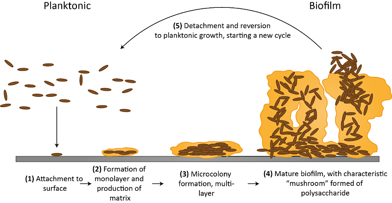
Biofilm formation can be divided into five stages: Initial reversible attachment (1), irreversible attachment (2-3), maturation (4) and dispersion (5) as shown in Figure 2. The initial contact of the moving planktonic bacteria with the surface is the starting point, which is still reversible at this stage. The bacteria will then start to form a monolayer and will produce an extracellular matrix or “slime” for protection. The matrix consists of extracellular polysaccharides, structural proteins, cell debris and nucleic acids; referred to as extracellular polymeric substances (EPS). The initial steps of the matrix formation are dominated by extracellular DNA (eDNA), whereas polysaccharides and structural proteins take over later on. In these stages, the formation of microcolonies takes place, which exhibit significant growth and cell-cell communication such as quorum sensing. The biofilm grows in a three-dimensional manner and the attachment is now irreversible. In the last stage, some cells of the mature biofilm start to detach and disperse into the environment as planktonic cells again to potentially start a new cycle of biofilm formation.
The role of biofilms in pathogenesis
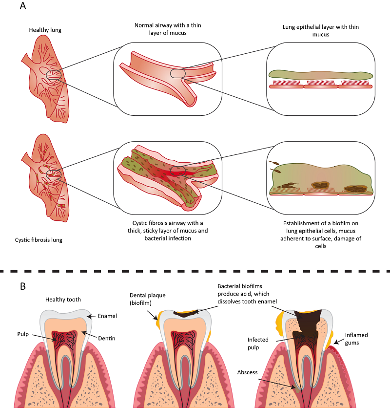
Biofilms can be found almost anywhere and may impact human health both positively and negatively. One example of a positive effect includes the biofilms of commensal bacteria such as Staphylococcus epidermidis, which can impede the colonisation of potentially pathogenic bacteria through the stimulation of host-cell immune defences and the prevention of adhesion. However, biofilms are more often associated with many pathogenic forms of human diseases and plant infections. One common example is cystic fibrosis, the most frequently passed genetic disorder in Western Europe. Cystic fibrosis (CF) patients suffer from chronic P. aeruginosa infections. When infecting the CF lung, P. aeruginosa undergoes a characteristic transition from an acute virulent pathogen to a CF‑adapted pathogen, allowing it to persist in the lung for years or even decades. This is due to the overproduction of the matrix polysaccharide alginate, leading to the formation of a mucoid biofilm that tolerates antibiotics, components of both the innate and adaptive immune response, and resists phagocytosis. The persistence of these mucoid biofilms within the CF lung leads to the development of a distinct antibody response. This prompts chronic inflammation mediated by granulocytes, and results in severe damage to the lung tissue of CF patients (see Figure 3 A). A second example for biofilms in human health is dental plaque potentially leading to dental caries. The consumption of fermentable carbohydrates such as sugary treats or drinks causes an increase in the production and secretion of organic acids by the bacteria found in dental plaque. If left untreated, the increased acidification of the biofilm leads to the demineralisation of the enamel and the formation of dental caries (see Figure 3 B).
Future directions
Due to the widespread distribution of biofilms in diseases and their resilience to numerous antimicrobial treatments, biofilm research is receiving more attention. Owing to increasing antimicrobial resistance, the focus of current research is shifting from targeting bacterial growth/division that causes cell death or dormancy, towards novel approaches. Examples include triggering the dispersal of the biofilm or looking into ways of preventing the initial formation, for instance by re-engineering the surfaces they are prone to develop upon such as urinary catheters and implants.